TREATMENTS - PLANTAR FASCIITIS
Heel Pain Syndrome
WHAT'S THE PROBLEM?
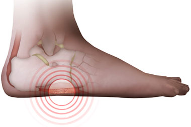
Plantar fasciitis/heel pain syndrome is an inflammation of a thick band of tissue at the bottom of the foot called the plantar fascia. The inflammation of the plantar fascia, at it's origin at the heel bone (calcaneus), causes the classic symptoms of pain at the bottom and/or side of the heel facing the opposite foot, often the most painful upon arising in the morning or when standing after sitting for prolonged periods. We call this post-static pain...pain after rest. This is because the plantar fascia is tight after rest, and the stretching inflames the painful area even more, therefore increasing the discomfort. The pain can exist with or WITHOUT a "heel spur". It is the feeling of most physicians that treat this condition often, that the pain is caused by the stretching and inflammation of the plantar fascia, and NOT by the bone spur. The spur is a result of pulling of the plantar fascia at the heel bone, resulting in proliferation of bone, often referred to as a "heel spur".
HOW DOES IT FEEL?
The classic symptoms are pain and a feeling of stiffness in the bottom and/or side of the heel. This pain is often a sharp pain that is described as a feeling of stepping on a stone or nail. The pain often reduces after a few steps, though it may still persist. This pain can also occur when walking after sitting for a prolonged time, such as sifting at work, driving a car, etc.
LET'S DO A TEST!
A thorough examination by a doctor of the clinical signs and symptoms will usually result in a clear diagnosis. X-rays will usually be taken to determine if any abnormality is present in the heel bone (calcaneus). On occasion, fractures, bone cysts, or foreign objects (pieces of metal, splinters, broken needles, glass) can be discovered as the cause of heel pain. If the presentation is not clear, other diagnostic tests may be utilized. Bone scans are often used if a bone cyst or abnormality in the bone are suspected or if a fracture is suspected. An MRI may be performed if a small "stress" type fracture is suspected. Additionally, an MRI is useful in determining the status of the surrounding tissue and can determine if there is a tear of the fascia or any other soft tissue and bone abnormalities. A CT scan is useful when suspecting a fracture of the heel bone and the extent of the fracture. Additionally, blood tests can be utilized to rule out arthritic or infectious disorders.
HOW DID THIS HAPPEN?
Plantar fasciitis heel pain syndrome can occur via a myriad of causes. Risk factors such as weight gain, foot type (flat feet, high arched feet), a high level of activity, sports, overuse, improper shoegear, improper support of the feet, trauma, tightness of muscles and daily activities can trigger the classic symptoms. Often, there is no single cause, but a culmination of several risk factors. It is imperative to address all of these factors to treat this condition successfully.
WHAT CAN I DO FOR IT?
Switching shoe gear to a well supportive running shoe can be effective. Reducing your level of activity helps. When you are off your feet, the injury is healing, it's getting better. When you are standing with inadequate foot support, it is getting injured. An orthotic or arch support can support your foot well enough to virtually eliminate the injury that is occurring while standing and walking.
WHAT WILL MY DOCTOR DO FOR IT?
Your podiatrist will perform a thorough examination and history of your condition. He/she will explain the cause of the problem and offer you various treatment options. X-rays will usually be taken to assess the status of the heel bone (calcaneus) and to check for other findings such as arthritis, foreign objects, fractures, etc. There are numerous treatment options, and the treatment protocol will vary according to the severity of the patient's symptoms.
Treatment options can include oral anti-inflammatory medications, heel cushions, heel cups, physical therapy, stretching exercises, taping/strapping of the foot, over-the-counter inserts, custom orthoses, injections, soft tissue wraps, weight loss, change of shoes, casting, night splints and surgery. Each option has advantages and disadvantages, and must be tailored to the needs and unique characteristics of each patient.
Conservative care is successful 85% of the time, and surgical treatment is usually performed 5-10% of the time. The surgical techniques differ according to the surgeon's preference. The surgery is usually performed as an outpatient and can be performed "traditionally", with removal of the spur and surgical release of the ligament, or via newer techniques utilizing endoscopic equipment. This newer technique utilizes two very small incisions and surgically releases a portion of the plantar fascia, and the "spur" is left undisturbed. The technique utilized should be discussed with the patient and will depend on x-ray and clinical findings.
CAN I PREVENT FROM IT HAPPENING AGAIN?
Controlling body weight, wearing supportive shoes and having additional support in your shoes can decrease the chance of recurrence. Additionally, stretching the Achilles Tendon and hamstrings and maintaining flexibility are extremely important in preventing recurrence of the problem. Most importantly, pain and discomfort are your body's signals that a problem exists. Prompt attention and treatment can often result in significant relief with the avoidance of complications.
|